59 Animal Cell Seen Under Electron Microscope
The electron microscope is more powerful than the light. All you need for this is a microscope with a basic transmitted light source and enough magnification to resolve individual yeast cells.

Electron Microscopic Study Of Cell And Organelles Important
A Based on the diagram state whether it represents an animal cell or plant cell b Give two reasons for your answer in a above c Why is the palisade layer a tissue.
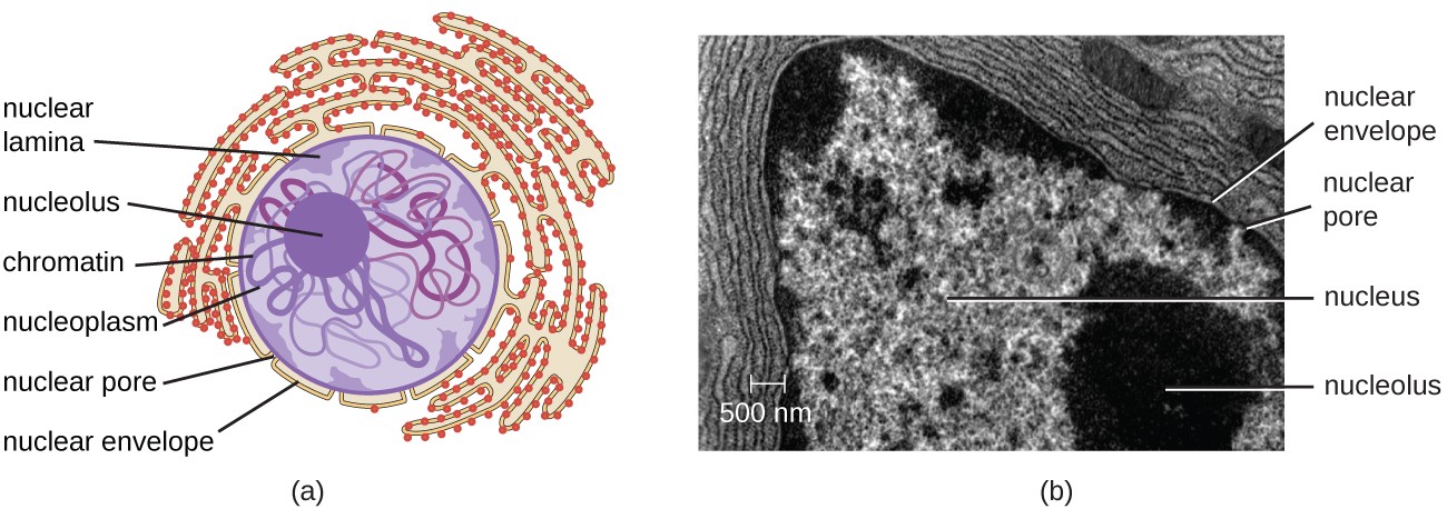
Animal cell seen under electron microscope. The cell membrane encloses the contents of the cell and separates it from its environment. It has small vacuoles. Electron microscope can magnify an object up to 500 000 times.
Here is an electron micrograph of an animal cell with the labels superimposed. They developed cell theory as a result of their studies. These are both specific types of.
Iii Mention any two structures found only in plant cell not in animal cell. Cells are microscopic and can only be seen under a microscope. Both the global and high-resolution distribution of colloidal gold labels on cells can be readily determined.
The cell wall nucleus vacuoles mitochondria endoplasmic reticulum Golgi apparatus and ribosomes are easily visible in this transmission electron micrograph. It also has a very high resolving power. Plant cell as shown above.
In the given figure of an animal cell as observed under an electron microscope. Unlike the eukaryotic cells of plants and fungi animal cells do not have a cell wall. This feature was lost in the distant past by the single-celled organisms that gave rise to the kingdom Animalia.
The diagram below represents a cell as seen under an electron microscope. I Name the parts labelled as 1 to 10. The Cell as Seen under the Electron Microscope.
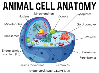
Typical Animal Cell Pinocytotic vesicle Lysosome Golgi vesicles Golgi vesicles rough ER endoplasmic reticulum Smooth ER no ribosomes Cell plasma membrane Mitochondrion Golgi apparatus Nucleolus Nucleus Centrioles 2 Each composed of 9 microtubule triplets Microtubules. Plant and animal cells have cell membranes cytoplasm a nucleus and organelles such as mitochondria and sometimes vacuoles. The high resolving power makes the electron microscope.
A cell is a very tiny structure which exists in living bodies. The plant cell as more rigid and stiff walls. Topic 1 2 Ultra Structure Of Cells Amazing World Of Science With.
Ii Which parts are concerned with the following functions. How is it different from animal cell. The animal cell is more fluid or elastic or malleable in structure.
It uses a beam of electrons to illuminate the specimen instead of light as in the case of light microscope. Electron Micrograph Animal Cell Under Electron Microscope. Below the basic structure is shown in the same animal cell on the left viewed with the light microscope and on the right with the transmission electron.
Most cells both animal and plant range in size between 1 and 100 micrometers and are thus visible only with the aid of a microscope. Animal cells have a basic structure. What Cell Organelles Can Be Seen Under The Electron Microscope But.
Visualization Of Plant Cell Wall Epitopes Using Immunogold. Illustrate only a plant cell as seen under electron microscope. A Release of energy b Protein synthesis c Transmission of hereditary characters from parents to their off springs.
Organelles which can be seen under light microscope are nucleus cytoplasm cell membrane chloroplasts and cell wall. Organelles which can be seen under electron microscope highest magnification to more than 200000x are ribosomes endoplasmic reticulum lysosomes centrioles. Some of the cell organelles that can be observed under the light microscope include the cell wall cell membrane cytoplasm nucleus vacuole and chloroplasts.
You see that many features are in common. The scientists Schleiden and Schwann observed plant and animal tissues under the microscope and both described similar cellular structures. Animal Cell as shown above.
Describe how turgor pressure builds up. I Name the parts labelled as 1 to 10. However no obvious structural damage.
This is the reason why you need to use a microscope to observe a cell. Asked Nov 28 2017 in Class IX Science by ashu Premium 930 points. The diagram below shows the general structure of an animal cell as seen under an electron microscope.
Imageanimal cell seen under Electron microscope ImagePlant cell seen under Electron microscope. Resolving power is the ability to distinguish between separate things which are close to each other. These cell organelles perform specific functions within the cell.
You know what the onion cells look like bricks of a parapet wall when you see it under the low power of microscope. You cant see any cell with your naked eye because they are very smaller than what human eyes can see normally. A typical animal cell as seen in an electron microscope Medical Images For PowerPoint.
In the given figure of an animal cell as observed under an electron microscope. What can be seen under a electron microscope. Under the intense radiation of the electron microscope 011 electron per Å 2 the question of viability of cells naturally arises because the amount of radiation absorbed during highmagnification imaging is sufficient to cause cell death.
95 Animal Cell Under Light Microscope Labelled
But at the same time it is interpretive. Dry the slide carefully with filter paperabsorbent paper and label it.
Unique Characteristics Of Eukaryotic Cells Microbiology
CZ02-003d Stomach - cat parietal cells with.
Animal cell under light microscope labelled. During mitosis the two sets of chromosomes are precisely separated. You see that many features are in common. Aims of the experiment.
Mitochondria - which can only just be seen using the light microscope - are the site of aerobic respiration which is the release of energy which powers the. Posted 6 years ago. The animal cell is more fluid or elastic or malleable in structure.
CZ01-008x Neurons - giant multi polar neurons. This appears at the light microscope level as a duplication of chromosomes. Result Conclusion SKILL ATTAINMENT PREPARE AND EXAMINE ONE ANIMAL CELL UNSTAINED AND STAINED USING THE LIGHT MICROSCOPE 100 400.
CZ02-003p Stomach - cat parietal cells with. Examine under the microscope following the usual procedure. Draw and label the structure of a generalized animal cell ie.
Similarly what does a animal cell look like under a microscope. Likewise can rough endoplasmic reticulum be seen. Under a microscope plant cells from the same source will have a uniform size and shape.
Before cell division the entire genome is copied. The diagram is very clear and labeled. We all do not forget that the human physique is quite problematic and a method I.
16 p4 which is photomicrograph of actual animal cells. This subject is important because in Biology we will be using the microscope many times during different laboratory exercises. Animal cells have a basic structure.
Labelled diagram of a plant cell under microscope posted on march 18 2011 by admin onion cells stained with methylene blue look at the images of onion cells as they would be seen under a microscope draw each magnification label appear high picture plant and animal cell. These cell organelles perform specific functions within the cell. Diagram Of Animal Cell Under Electron Microscope.
Ribosomes are only visible with an electron. This shows a generalized animal cell under a light microscope. A brief explanation of the.
Monday April 5th 2021. CZ08-001c Scalp - old skin flaking off hair. Below the basic structure is shown in the same animal cell on the left viewed with the light microscope and on the right with the transmission electron.
The plant cell as more rigid and stiff walls. Some of the cell organelles that can be observed under the light microscope include the cell wall cell membrane cytoplasm nucleus vacuole and chloroplasts. Diagram Of Animal Cell Under Electron Microscope Labeled.
To use a light microscope to examine animal or plant. 14 can you distinguish features of the cells in Fig. Animal Plant Cells Gcse Science Biology Get To Know Science Youtube Mitochondrion are visible with a light microscope but cant be seen in detail.
Viewing Animal Cells under a microscope. Animal Cell as shown above Plant cell as shown above. Photo Album By Darcy Plant And Animal Cells Under The Microscope.
Ziehen die pins an die richtige stelle auf dem bild. Introduction The purpose of this lab was to use the microscope and identify cells such as animal cells and plant cells. Once slides have been prepared they can be examined under a microscope.
Draw labelled diagrams of what you see at 100 and at 400. Microscopic Animal Cells 82 images View. Module 5 Page 2.
Investigating cells with a light microscope. Here is an electron micrograph of an animal cell with the labels superimposed. Using the labels of Fig.
Beneath a plant cells cell wall is a cell membrane. Structure Of Nucleolus Under Light And Electron Microscope A. These are both specific types of.
Its a thin slice. Animal and plant cells undergo a precise type of division called mitosis. Labelled animal cell diagram gcse.
Heres a diagram of a plant cell. The microscope is used for looking at many specimens that cannot be seen with the. Illustrate Only A Plant Cell As Seen Under Electron Microscope.
In most plant cells the organelles that are visible under a compound light microscope are the cell wall cell membrane cytoplasm central vacuole and nucleus. So lets begin by drawing a rough-oval shape. Microscopically animal cells from the same tissue of an animal will have varied sizes and shapes due to the lack of a rigid cell wall.
CZ07-003c Skeletal Muscle - striated muscle. Plant Cell Under Microscope Labeled.
