74 Animal Cell Diagram Not Labeled
Quiz by SnelsonBiology. This will help you understand how much you have comprehended.
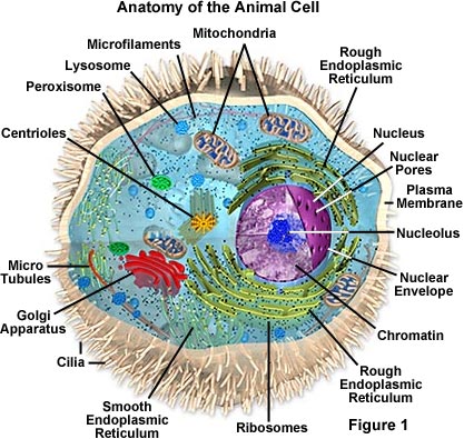
Molecular Expressions Cell Biology Animal Cell Structure
Colors Venn Diagram 175.

Animal cell diagram not labeled. Planets of the Solar System Venn Diagram 156. 25 Blank Plant Cell Diagram Worksheet. Animal-cell-diagram-not-labeled Tims Printables Mathilda Barnes The significant differences between plant and animal cells are also shown and the diagrams are followed by more in-depth information.
Animal cell diagram not labeled. Simple black and white doodle of pedestrians. Cells are made up of different parts.
Can you label the Animal cell. Lets find out how many you get right. The nucleus regulates cell.
There are various organelles present within the cell and are classified into three categories based on the presence or absence of membrane. Labeled diagram of mitosis of an animal cell. The following animal cell diagram labeled show more parts of the cell.
Test your knowledge on this science quiz and compare your score to others. The following animal cell diagram labeled show more parts of the cell. A labeled diagram of the plant cell and functions of its organelles we are aware that all life stems from a single cell and that the cell is the most basic unit of all living organisms.
Animal Cell Diagram Not Labeled. In a typical animal cell mitosis. The various cell organelles present in an animal cell are clearly marked in the animal cell diagram provided below.
5th grade science and biology. The Cell Organelles are membrane-bound present within the cells. Animal cells do not have cell walls.
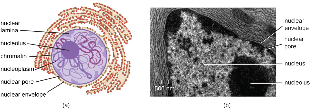
Nucleus The cell nucleus is an organelle that contains most of the cells genetic material. As you read the information on each organelle refer to the animal cell diagram for better clarity. Unlabeled Animal Cell Diagram.
The important part is that it does not have any sharp edges. Listed below are the Cell Organelles of an animal cell along with their functions. Interphase the cell when not undergoing mitosis but the dna is replicated prophase metaphase anaphase and.
This may be useful as a printable poster for the classroom or as part of a presentation or report. Science Diagrams Below you. Printable Animal Cell Diagram Labeled Unlabeled And Blank Finally an unlabeled version of the diagram is included at the bottom of the page in color and black and white.
Science Printables Read More Article by Tims Printables. Animal cell diagram detailing the various organelles Though this animal cell diagram is not representative of any one particular type of cell it provides insight into the primary organelles and the intricate internal structure of. Well-Labelled Diagram of Animal Cell.
Can you label the Animal cell. Printable Animal Cell Diagram Labeled Unlabeled and Blank. Printable animal cell diagram to help you learn the organelles in an animal cell in preparation for your test or quiz.
Plant and animal cell diagram not labeled. Free UK Delivery on Eligible Orders Labellers Label Applicators and Label Print Apply from ALTech UK Well-Labelled Diagram of Animal Cell The Cell Organelles are membrane-bound. Have chloroplasts and use photosynthesis to produce food have cell wall made of cellulose a plant cell has plasmodesmata microscopic channels which traverse the cell walls of the cells one very large vacuole in the center are rectangular in shape animal cells.
Plant Cell Project Models Plant Cell Model Plant Cell Drawing Animal Cell Structure Plant Cell Diagram Science Diagrams Plant And Animal Cells Cell Wall Science Projects. Feb 28 2020 - Blank Plant Cell Diagram Worksheet. The animal cell diagram is widely asked in class 10 and 12 examinations and is beneficial to understand the structure.
Printable animal cell diagram labeled unlabeled and blank. A bacteria diagram clearly helps us to profit extra approximately this unmarried cell organisms which have neither membrane-bounded nucleolus or organelles like mitochondria and chloroplasts. In this video Im going to draw labelled diagram of animal cellin this video you will see the diagram of Animal cell and its labellingThis diagram of.
Learning about animal cells can be tricky at first so why dont we start you off relatively easy with this animal cell part labelling quiz. Blank Animal Cell Diagram Worksheet Driverlayer Search. Plant Cell And Animal Cell Diagram 8th Standard.
Difference between mitosis and meiosis. To check if you have understood the cell parts draw a blank animal cell diagram and try to fill in the different parts without referring to the labeled one given here. Mitosis is a process of cell division which results in the production of two daughter cells from a single parent cell.
The structure labeled G give rise to spindle fibers and exclusively seen in animal cell. Organelles and their Functions. Sep 12 2020 usher.
Which of the following organelles does an animal cell NOT have. Cells are made up of different parts. In this one well be giving you a question referring to a given diagram and asking you to label it.
Article by Tims Printables. Prove You Arent a Robot - Animal Camouflage V 155. Finally an unlabeled version of the diagram is included at the bottom of the page in color and black and white.
95 Animal Cell Under Light Microscope Labelled
But at the same time it is interpretive. Dry the slide carefully with filter paperabsorbent paper and label it.
Unique Characteristics Of Eukaryotic Cells Microbiology
CZ02-003d Stomach - cat parietal cells with.
Animal cell under light microscope labelled. During mitosis the two sets of chromosomes are precisely separated. You see that many features are in common. Aims of the experiment.
Mitochondria - which can only just be seen using the light microscope - are the site of aerobic respiration which is the release of energy which powers the. Posted 6 years ago. The animal cell is more fluid or elastic or malleable in structure.
CZ01-008x Neurons - giant multi polar neurons. This appears at the light microscope level as a duplication of chromosomes. Result Conclusion SKILL ATTAINMENT PREPARE AND EXAMINE ONE ANIMAL CELL UNSTAINED AND STAINED USING THE LIGHT MICROSCOPE 100 400.
CZ02-003p Stomach - cat parietal cells with. Examine under the microscope following the usual procedure. Draw and label the structure of a generalized animal cell ie.
Similarly what does a animal cell look like under a microscope. Likewise can rough endoplasmic reticulum be seen. Under a microscope plant cells from the same source will have a uniform size and shape.
Before cell division the entire genome is copied. The diagram is very clear and labeled. We all do not forget that the human physique is quite problematic and a method I.
16 p4 which is photomicrograph of actual animal cells. This subject is important because in Biology we will be using the microscope many times during different laboratory exercises. Animal cells have a basic structure.
Labelled diagram of a plant cell under microscope posted on march 18 2011 by admin onion cells stained with methylene blue look at the images of onion cells as they would be seen under a microscope draw each magnification label appear high picture plant and animal cell. These cell organelles perform specific functions within the cell. Diagram Of Animal Cell Under Electron Microscope.
Ribosomes are only visible with an electron. This shows a generalized animal cell under a light microscope. A brief explanation of the.
Monday April 5th 2021. CZ08-001c Scalp - old skin flaking off hair. Below the basic structure is shown in the same animal cell on the left viewed with the light microscope and on the right with the transmission electron.
The plant cell as more rigid and stiff walls. Some of the cell organelles that can be observed under the light microscope include the cell wall cell membrane cytoplasm nucleus vacuole and chloroplasts. Diagram Of Animal Cell Under Electron Microscope Labeled.
To use a light microscope to examine animal or plant. 14 can you distinguish features of the cells in Fig. Animal Plant Cells Gcse Science Biology Get To Know Science Youtube Mitochondrion are visible with a light microscope but cant be seen in detail.
Viewing Animal Cells under a microscope. Animal Cell as shown above Plant cell as shown above. Photo Album By Darcy Plant And Animal Cells Under The Microscope.
Ziehen die pins an die richtige stelle auf dem bild. Introduction The purpose of this lab was to use the microscope and identify cells such as animal cells and plant cells. Once slides have been prepared they can be examined under a microscope.
Draw labelled diagrams of what you see at 100 and at 400. Microscopic Animal Cells 82 images View. Module 5 Page 2.
Investigating cells with a light microscope. Here is an electron micrograph of an animal cell with the labels superimposed. Using the labels of Fig.
Beneath a plant cells cell wall is a cell membrane. Structure Of Nucleolus Under Light And Electron Microscope A. These are both specific types of.
Its a thin slice. Animal and plant cells undergo a precise type of division called mitosis. Labelled animal cell diagram gcse.
Heres a diagram of a plant cell. The microscope is used for looking at many specimens that cannot be seen with the. Illustrate Only A Plant Cell As Seen Under Electron Microscope.
In most plant cells the organelles that are visible under a compound light microscope are the cell wall cell membrane cytoplasm central vacuole and nucleus. So lets begin by drawing a rough-oval shape. Microscopically animal cells from the same tissue of an animal will have varied sizes and shapes due to the lack of a rigid cell wall.
CZ07-003c Skeletal Muscle - striated muscle. Plant Cell Under Microscope Labeled.
59 Animal Cell Seen Under Electron Microscope
The electron microscope is more powerful than the light. All you need for this is a microscope with a basic transmitted light source and enough magnification to resolve individual yeast cells.

Electron Microscopic Study Of Cell And Organelles Important
A Based on the diagram state whether it represents an animal cell or plant cell b Give two reasons for your answer in a above c Why is the palisade layer a tissue.
Animal cell seen under electron microscope. The cell membrane encloses the contents of the cell and separates it from its environment. It has small vacuoles. Electron microscope can magnify an object up to 500 000 times.
Here is an electron micrograph of an animal cell with the labels superimposed. They developed cell theory as a result of their studies. These are both specific types of.
Iii Mention any two structures found only in plant cell not in animal cell. Cells are microscopic and can only be seen under a microscope. Both the global and high-resolution distribution of colloidal gold labels on cells can be readily determined.
The cell wall nucleus vacuoles mitochondria endoplasmic reticulum Golgi apparatus and ribosomes are easily visible in this transmission electron micrograph. It also has a very high resolving power. Plant cell as shown above.
In the given figure of an animal cell as observed under an electron microscope. Unlike the eukaryotic cells of plants and fungi animal cells do not have a cell wall. This feature was lost in the distant past by the single-celled organisms that gave rise to the kingdom Animalia.
The diagram below represents a cell as seen under an electron microscope. I Name the parts labelled as 1 to 10. The Cell as Seen under the Electron Microscope.
Typical Animal Cell Pinocytotic vesicle Lysosome Golgi vesicles Golgi vesicles rough ER endoplasmic reticulum Smooth ER no ribosomes Cell plasma membrane Mitochondrion Golgi apparatus Nucleolus Nucleus Centrioles 2 Each composed of 9 microtubule triplets Microtubules. Plant and animal cells have cell membranes cytoplasm a nucleus and organelles such as mitochondria and sometimes vacuoles. The high resolving power makes the electron microscope.
A cell is a very tiny structure which exists in living bodies. The plant cell as more rigid and stiff walls. Topic 1 2 Ultra Structure Of Cells Amazing World Of Science With.
Ii Which parts are concerned with the following functions. How is it different from animal cell. The animal cell is more fluid or elastic or malleable in structure.
It uses a beam of electrons to illuminate the specimen instead of light as in the case of light microscope. Electron Micrograph Animal Cell Under Electron Microscope. Below the basic structure is shown in the same animal cell on the left viewed with the light microscope and on the right with the transmission electron.
Most cells both animal and plant range in size between 1 and 100 micrometers and are thus visible only with the aid of a microscope. Animal cells have a basic structure. What Cell Organelles Can Be Seen Under The Electron Microscope But.
Visualization Of Plant Cell Wall Epitopes Using Immunogold. Illustrate only a plant cell as seen under electron microscope. A Release of energy b Protein synthesis c Transmission of hereditary characters from parents to their off springs.
Organelles which can be seen under light microscope are nucleus cytoplasm cell membrane chloroplasts and cell wall. Organelles which can be seen under electron microscope highest magnification to more than 200000x are ribosomes endoplasmic reticulum lysosomes centrioles. Some of the cell organelles that can be observed under the light microscope include the cell wall cell membrane cytoplasm nucleus vacuole and chloroplasts.
You see that many features are in common. The scientists Schleiden and Schwann observed plant and animal tissues under the microscope and both described similar cellular structures. Animal Cell as shown above.
Describe how turgor pressure builds up. I Name the parts labelled as 1 to 10. However no obvious structural damage.
This is the reason why you need to use a microscope to observe a cell. Asked Nov 28 2017 in Class IX Science by ashu Premium 930 points. The diagram below shows the general structure of an animal cell as seen under an electron microscope.
Imageanimal cell seen under Electron microscope ImagePlant cell seen under Electron microscope. Resolving power is the ability to distinguish between separate things which are close to each other. These cell organelles perform specific functions within the cell.
You know what the onion cells look like bricks of a parapet wall when you see it under the low power of microscope. You cant see any cell with your naked eye because they are very smaller than what human eyes can see normally. A typical animal cell as seen in an electron microscope Medical Images For PowerPoint.
In the given figure of an animal cell as observed under an electron microscope. What can be seen under a electron microscope. Under the intense radiation of the electron microscope 011 electron per Å 2 the question of viability of cells naturally arises because the amount of radiation absorbed during highmagnification imaging is sufficient to cause cell death.
88 Animal Vs Plant Cell Differences
Plant cells have a cell wall surrounding cell membrane whereas animal cells only have a cell membrane. Beyond size the main structural differences between plant and animal cells lie in a few additional structures found in plant cells.
What Is The Difference Between Plant Cell Vacuoles And Animal Cell Vacuoles Quora
Animal cells do not have a cell wall.

Animal vs plant cell differences. Thus in plants the gametes are formed by mitosis not meiosis which. Plant cells can be larger than animal cells. Animal cells contain lysosomes which are absent in plant cells.
The mode of cytokinesis is one of the most critical features that differentiate the mechanism of the cell division in the plant and animal cell. As plants and animals are eukaryotic so they have almost similar cellular structure but few organelles like chloroplast plasmodesmata cell wall plastids etc. The processes are quiet the same in plants and animals.
Animal cells are generally smaller than plant cells. As a result most animal cells are round and flexible whereas most plant cells are rectangular and rigid. Instead they have cilia the.
Differences Between Animal Cells and Plant Cells Size. It is obvious why animal cells lack in chloroplast as there is no process of photosynthesis that generates food for the cells - animal cells create their energy via different process.
18 rows A difference between plant cells and animal cells is that most animal cells are round. However in animals it results into the formation of gametes which is a reproductive or sex cell. Animal cells do not have a cell wall or chloroplasts but plant cells do.
Plant cells are larger than animal cells. Because of functional differences there are great differences between plant and animal cells. Animal cells and plant cells share the common components of a nucleus cytoplasm mitochondria and a cell membrane.
G PC and AC explore the differences similarities between a plant cell versus an animal cell in Mrs. A difference between plant cells and animal cells is that plant cells have a rigid cell wall that surrounds the cell membrane. The size of animal cells can range from 10 to 30 micrometers.
Animal cells have one or more small vacuoles but plant cells have only one big vacuole. Animal cells range from 10 to 30 micrometers in length while. Plant cells have three extra components a vacuole chloroplast and a cell wall.
Come along as Mrs. The primary difference from plant cells is that animal cells dont contain chloroplast nor structurally important cell walls. The normal range for an animal cell varies from 10 to 30 micrometers while that for a plant cell stretches from 10 to 100 micrometers.
A plant cell produces daughter cells by the cell-constriction mechanism or through the formation of cell-furrow. The normal range of the animal. On the other hand in plants it forms spores which further grow into gametophyte.
Are only found in the plant cell while in there is no cell wall in the animal cell. Plant cells have chloroplasts that help in photosynthesis. 29 rows The plant cell can also be larger than the animal cell.
These are absent in animal cells. One example of this is that plant cells have chloroplasts that allow them to perform photosynthesis for energy but animal cells do not have chloroplasts since they. The normal range for a plant cell is between 10 to 100 micrometers.
Animal cells come in various sizes and tend to have round or irregular shapes. Animal cells are round and irregular in shape but plants cells are rectangular and have a fixed shape. Apart from size there are several other structural differences between plant and animal cells too.
Meiosis is a type of cell division which reduces the chromosome number. 9 rows Unlike animal cells plant cells have cell walls and organelles called chloroplasts. The key difference between plant cell and animal cell is the presence and absence of.
67 Animal Cell Structure And Function Masteringbiology
Week 2 - Chapter 4 Laboratory. Learn vocabulary terms and more with flashcards games and other study tools.

Prokaryotic Cell Structure And Function Masteringbiology Biology A Global Approach Plus Masteringbiology With Pearson Etext Global Edition
Which of these organelles carries out cellular respiration.

Animal cell structure and function masteringbiology. Lets begin with the components of the animal cells-Cell membrane. Nucleic acid saturated fat unsaturated fat steroid protein Correct The fatty acid tails lack double bonds. Complex structures of large and small subunits each of which contains RNA and protein molecules.
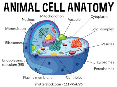
Plant cells and animal cells share some common features as both are eukaryotic cells. Animal cell diagram. Re beginning this tutorial watch the Tour of an Animal Cell animation to learn about the general structure of an animal cell.
These cells differ in their shapes sizes and their structure as they have to fulfil specific functions. 7 Cell Structure and Function. The membrane has the following functions-It encloses the various cell structure and cytoplasm.
Part A This animation illustrates the functioning of a _____ protein. Regulates cytoplasm composition creates internal pressure and stores cell compounds. Makes sugar by converting light energy into chemical energy.
Animal cells vary in different shapes and size and perform specific functions. They have a distinct nucleus with all cellular organelles enclosed in a membrane and thus called a eukaryotic cell. Appendage that propels the cell.
It regulates the movement of substance into and from the cell. Large organelle the contains most of the genes that control the cell. Part B Which of the following statements about monosaccharide structure is true.
The nucleus contains most of a cells DNA. Answer Correct Phospholipids are. Transport enzyme signal structural receptor Correct.
Receptor contractile structural protein gene regulatory transport Correct The protein is transporting a substance across the cell membrane. Learn vocabulary terms and more with flashcards games and other study tools. Wax cholesterol phospholipid RNA steroids Correct RNA is a nucleic acid Part B This figure is an example of an _____.
The cell is the structural and functional unit of life. Then click on the image to start the animation. Cell Structure and Function MasteringBiology- Pearson.
An animal cell is defined as the basic structural and functional unit of life in organisms of the kingdom Animalia. Part A Which molecule is not a carbohydrate. Animal Cell Structure and Function Part A.
Double membrane that encloses the nucleus. Start studying Chapter 6. Starch Glycogen Lipid Cellulose Correct A lipid is a hydrophobic polymer not a carbohydrate.
To understand how an organism works you must first understand how its cells are structured me roles that each structure performs in the cell. Start studying Mastering Biology HW 3 Animal Cell StructureFunction. Modifies and packages proteins.
Each one functions as a protein manufacturer and there may be 10000 in a single cell. 40 The Animal Body. 41 Chemical Signals in Animals.
UNIT 2 CELL BIOLOGY. In addition they have locomotory and cytoskeletal structures. They are considered to be multicellular organisms.
Animal cells have an organized nucleus with the nuclear envelope. View Homework Help - MasteringBiology_ Week 2 - Chapter 4 Laboratory Homework P3pdf from BIOS 140 at DeVry University Columbus North. Aldoses and ketoses differ in the position of their hydroxyl groups.
A group of cells assemble together to form tissues and eventually to organs and organ systems. Found throughout the cell interior. A six-carbon sugar is called a pentose.
Animal cells have centrioles which are absent in plant cells. To view the animation click here. Cell membrane cell boundary controls movement of materials in out recognizes signals cytoplasm jelly-like material holding organelles in place mitochondria make ATP energy from sugar O 2 nucleus protects DNA controls cell ribosomes builds proteins ER helps finish proteins makes membranes Golgi apparatus finishes packages ships proteins lysosome food digestion garbage disposal.
Structures and Functions BioFlix tutorial 6 of 8 ell is the basic structural and functional unit of all organisms. A Global Approach with MasteringBiology Global Edition 10th Edition. However they differ as animals need to adapt to a more active and non-sedentary lifestyle.
Part A Which of these is NOT a lipid. A cell is always surrounded by a thin membrane called plasma membrane. Structure and Characteristics of an Animal Cell.
Part C Which of these is a phospholipid. It gives definite shape to the cell. All monosaccharides contain carbon hydrogen oxygen and.
Choose the letter that indicates the organelle that contains most of a cells DNA. Produces chemical energy ATP that can power the cell. Part B Arrow A is indicating an _____ protein.
Strands containing DNA genes and associative protein.
